Scientific Image Gallery
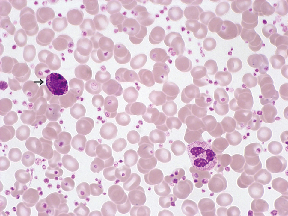
Peripheral blood (May-Grünwald-Giemsa stain) of a 27-year old patient with chronic myelogenous leukaemia (CML). Normal red blood cells, marked thrombocytosis (2,750,000/µL), increased basophilic granulocytes (->) and a normal white blood cell count are observed. In this case no JAK2 mutation could be detected; instead the patient was tested positive for the BCR-ABL fusion gene.
<p>Peripheral blood (May-Grünwald-Giemsa stain) of a 27-year old patient with chronic myelogenous leukaemia (CML). Normal red blood cells, marked thrombocytosis (2,750,000/µL), increased basophilic granulocytes (->) and a normal white blood cell count are observed. In this case no JAK2 mutation could be detected; instead the patient was tested positive for the BCR-ABL fusion gene.</p>
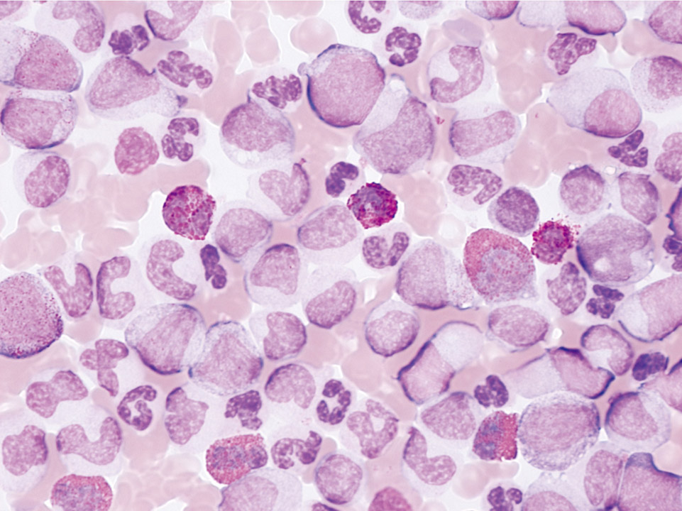
The peripheral blood (May-Grünwald-Giemsa stain) of a 27-year old patient with hearing loss showed a massive leukocytosis (680,000/µL) and a marked left shift with a distinct fraction of blast cells. Several eosinophilic cells are clearly visible. Platelets are abundant, although not visible in this part of the blood film. The BCR-ABL fusion gene was detected by FISH analysis of the peripheral blood and secured the diagnosis of CML. Because of the threat of permanent loss of hearing chemotherapy was started within hours after venepuncture (without waiting for the results from bone marrow cytology).
<p>The peripheral blood (May-Grünwald-Giemsa stain) of a 27-year old patient with hearing loss showed a massive leukocytosis (680,000/µL) and a marked left shift with a distinct fraction of blast cells. Several eosinophilic cells are clearly visible. Platelets are abundant, although not visible in this part of the blood film. The BCR-ABL fusion gene was detected by FISH analysis of the peripheral blood and secured the diagnosis of CML. Because of the threat of permanent loss of hearing chemotherapy was started within hours after venepuncture (without waiting for the results from bone marrow cytology).</p>
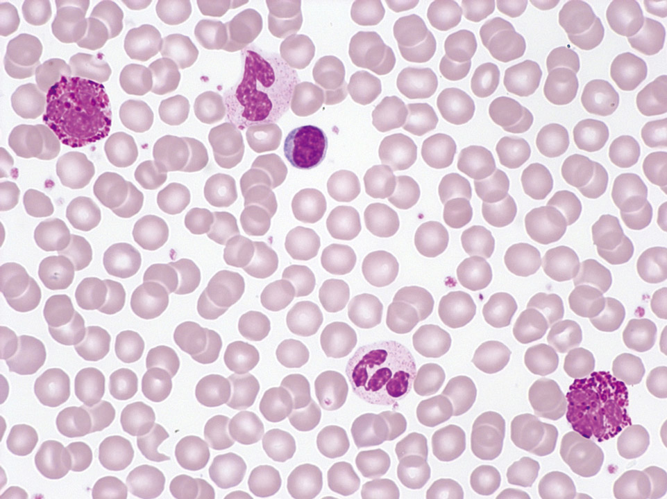
The peripheral blood (May-Grünwald-Giemsa stain) of a patient shows as an incidental finding leukocytosis with left shift up to myelocytes, basophilia and mild thrombocytosis. CML was suspected and later confirmed by cytogenetic demonstration of the Ph1.
<p>The peripheral blood (May-Grünwald-Giemsa stain) of a patient shows as an incidental finding leukocytosis with left shift up to myelocytes, basophilia and mild thrombocytosis. CML was suspected and later confirmed by cytogenetic demonstration of the Ph1.</p>
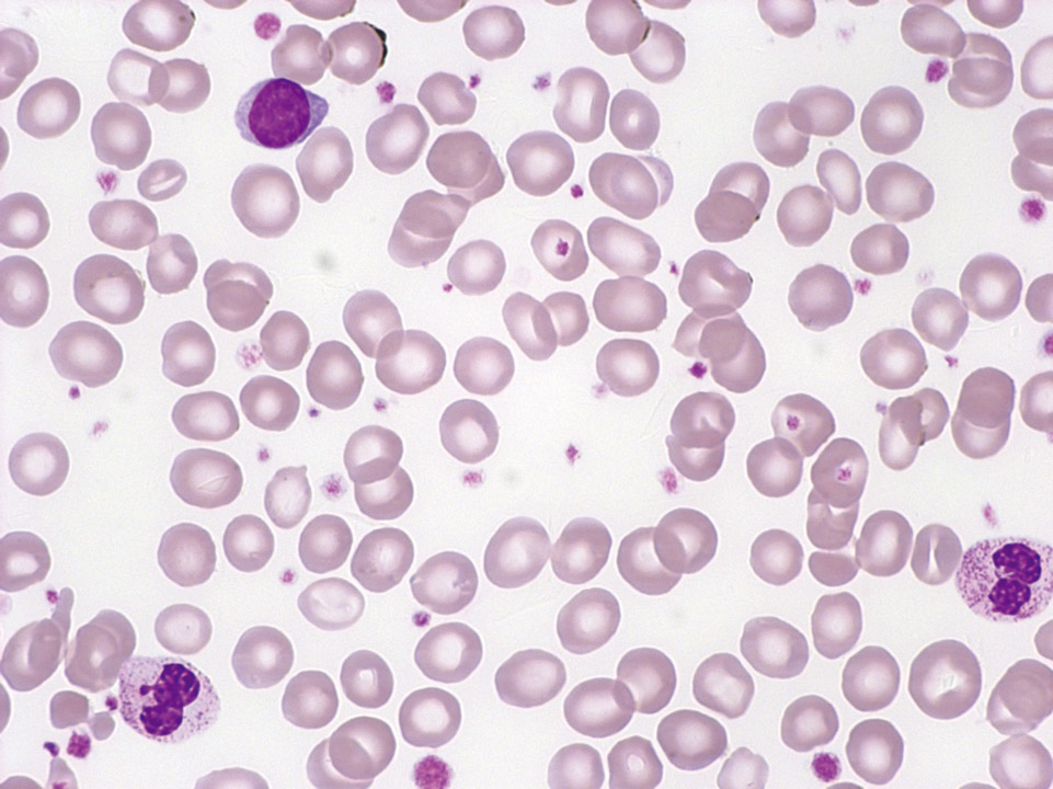
Peripheral blood (May-Grünwald-Giemsa stain) of a patient with ET showing an isolated thrombocytosis. A JAK2 mutation was detected by molecular biological techniques. The BCR-ABL fusion gene was not found.
<p>Peripheral blood (May-Grünwald-Giemsa stain) of a patient with ET showing an isolated thrombocytosis. A JAK2 mutation was detected by molecular biological techniques. The BCR-ABL fusion gene was not found.</p>
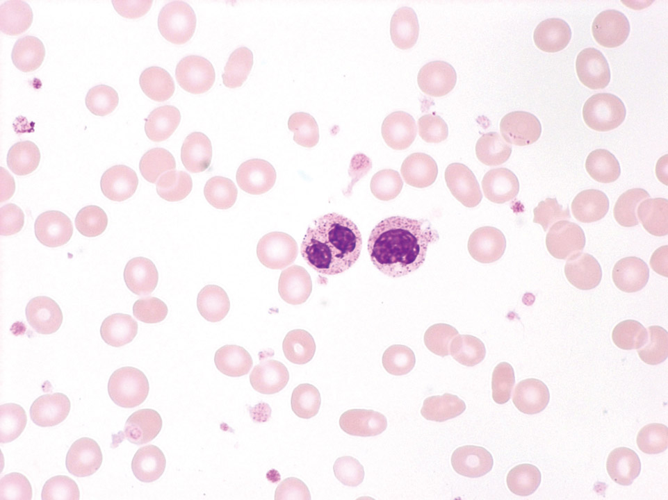
Peripheral blood (May-Grünwald-Giemsa stain) of a patient with MDS: on the left a pseudo-Pelger-Huet cell, on the right a dysplastic metamyelocyte. Anaemia and atypical platelets are also present. After peripheral blood and bone marrow analysis the diagnosis of refractory anaemia with excess of blasts (RAEB-1) was made.
<p>Peripheral blood (May-Grünwald-Giemsa stain) of a patient with MDS: on the left a pseudo-Pelger-Huet cell, on the right a dysplastic metamyelocyte. Anaemia and atypical platelets are also present. After peripheral blood and bone marrow analysis the diagnosis of refractory anaemia with excess of blasts (RAEB-1) was made.</p>
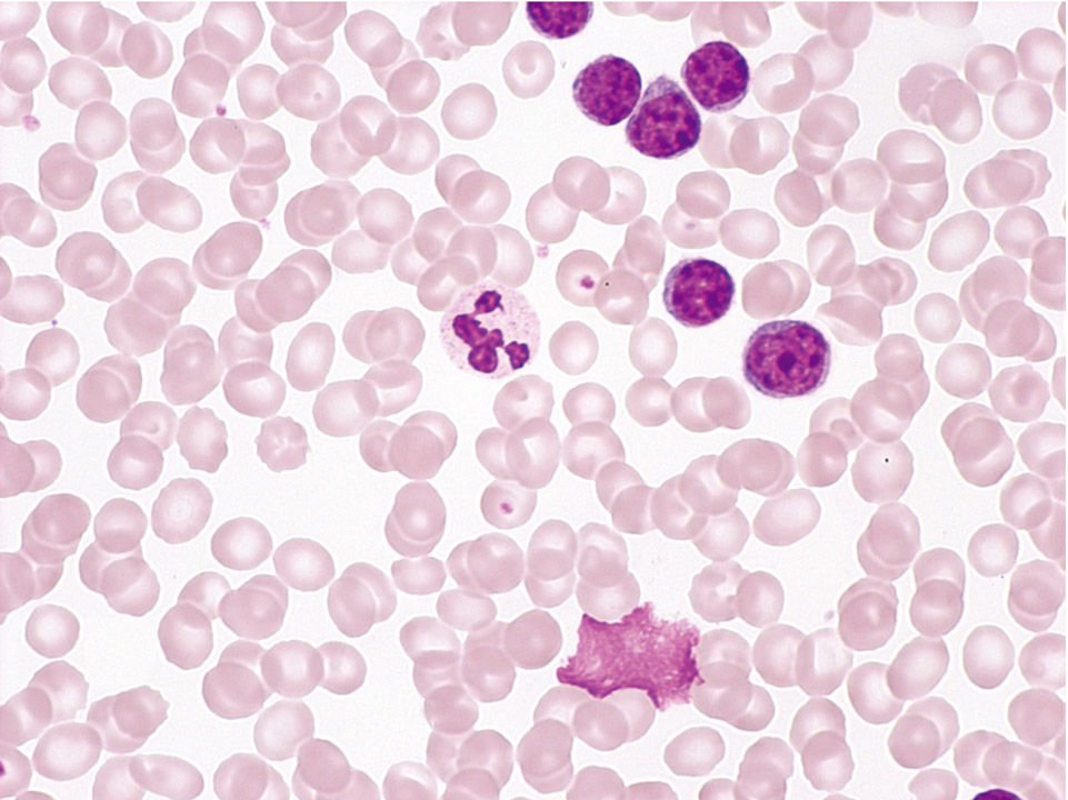
Peripheral blood (May-Grünwald-Giemsa stain) showing a typical B-CLL with an elevated lymphocyte count, normal granulocyte and platelet counts and normal red blood cells.
<p>Peripheral blood (May-Grünwald-Giemsa stain) showing a typical B-CLL with an elevated lymphocyte count, normal granulocyte and platelet counts and normal red blood cells.</p>
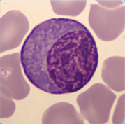
Cell description:
Size: up to 20 µm
Nucleus: eccentric with coarsely clumped chromatin, often clock-face chromatin pattern
Cytoplasm: strongly basophilic cytoplasm with apparent less basophilic Golgi zone adjacent to the nucleus
<p>Cell description: </p> <p>Size: up to 20 µm </p> <p>Nucleus: eccentric with coarsely clumped chromatin, often clock-face chromatin pattern </p> <p>Cytoplasm: strongly basophilic cytoplasm with apparent less basophilic Golgi zone adjacent to the nucleus</p>
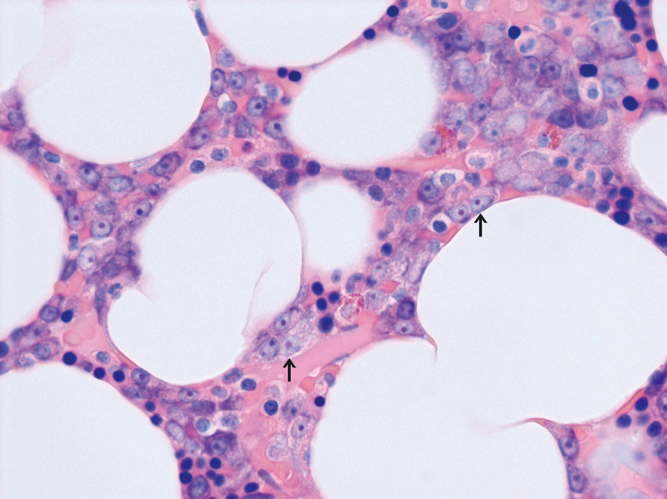
Bone marrow histology (Giemsa stain) of a patient with multiple myeloma showing groups (nests) of plasma cells (->) in atypical locations. Normal plasma cells are usually located close to the blood vessels.
<p>Bone marrow histology (Giemsa stain) of a patient with multiple myeloma showing groups (nests) of plasma cells (->) in atypical locations. Normal plasma cells are usually located close to the blood vessels.</p>
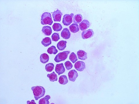
Plasma cells in a cytospin prepared from cerebrospinal fluid (CSF) of the patient with multiple myeloma, May-Gruenwald Giemsa stain
<p>Plasma cells in a cytospin prepared from cerebrospinal fluid (CSF) of the patient with multiple myeloma, May-Gruenwald Giemsa stain</p>


