Scientific Image Gallery
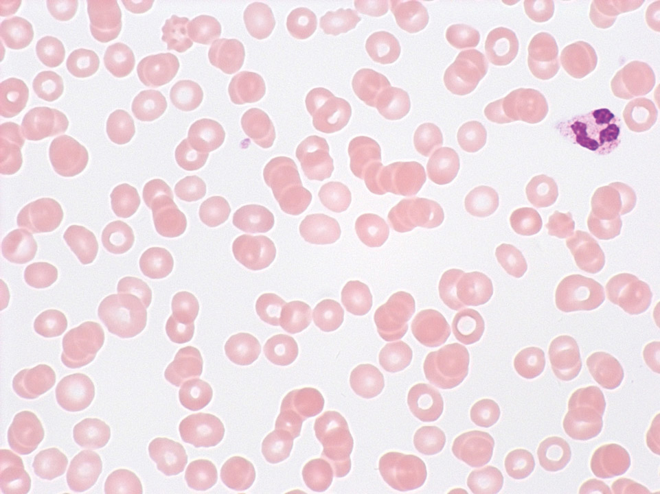
In severe anaemia (here severe aplastic anaemia with a haemoglobin concentration of 4.8 g/dL) or in erythrocytosis with a haematocrit above 50% it is difficult to prepare a proper blood film, with the blood film being either too thin (anaemia) or too thick. In cases of erythrocytosis it might be necessary to dilute the blood sample beforehand.
<p>In severe anaemia (here severe aplastic anaemia with a haemoglobin concentration of 4.8 g/dL) or in erythrocytosis with a haematocrit above 50% it is difficult to prepare a proper blood film, with the blood film being either too thin (anaemia) or too thick. In cases of erythrocytosis it might be necessary to dilute the blood sample beforehand.</p>
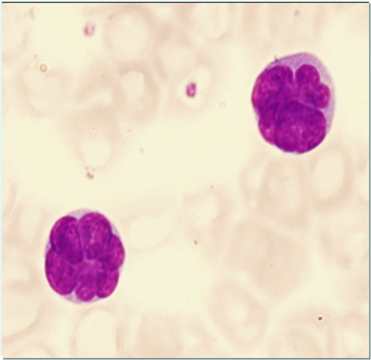
Typical for a Sézary cell is its cerebriform shape of the nucleus. The cytoplasm is usually basophilic with a medium nucleocytoplasmic ratio.
<p>Typical for a Sézary cell is its cerebriform shape of the nucleus. The cytoplasm is usually basophilic with a medium nucleocytoplasmic ratio. </p>
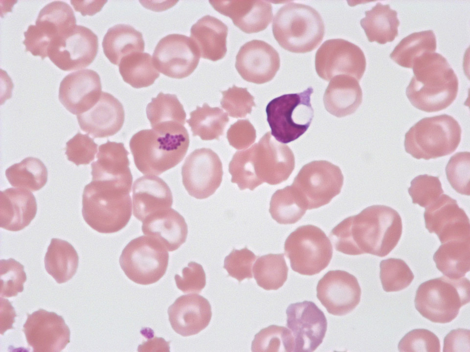
Accumulation of streptococci on a red blood cell of a patient in intensive care suffering from streptococcal septicaemia (further to the right, a lymphocyte with a cytoplasm vacuole can be seen).
<p>Accumulation of streptococci on a red blood cell of a patient in intensive care suffering from streptococcal septicaemia (further to the right, a lymphocyte with a cytoplasm vacuole can be seen).</p>
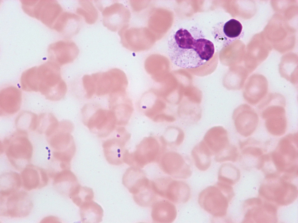
Streptococci, extracellular as well as intracellular (->), in a patient with finger gangrene. The patient died hours later despite intensive care treatment. If bacteria are detectable in a blood film, a blood film from a different patient should be stained with the same staining solution and be checked for bacteria to rule out contamination of the staining solution. If none are detectable, the detection of bacteria has to be reported to the treating physician immediately.
<p>Streptococci, extracellular as well as intracellular (->), in a patient with finger gangrene. The patient died hours later despite intensive care treatment. If bacteria are detectable in a blood film, a blood film from a different patient should be stained with the same staining solution and be checked for bacteria to rule out contamination of the staining solution. If none are detectable, the detection of bacteria has to be reported to the treating physician immediately.</p>
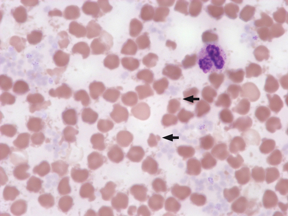
Striations with a blue cast and parts of the red blood cells broken off, due to overheating of the blood sample (storage in the car with the sun shining on it).
<p>Striations with a blue cast and parts of the red blood cells broken off, due to overheating of the blood sample (storage in the car with the sun shining on it).</p>
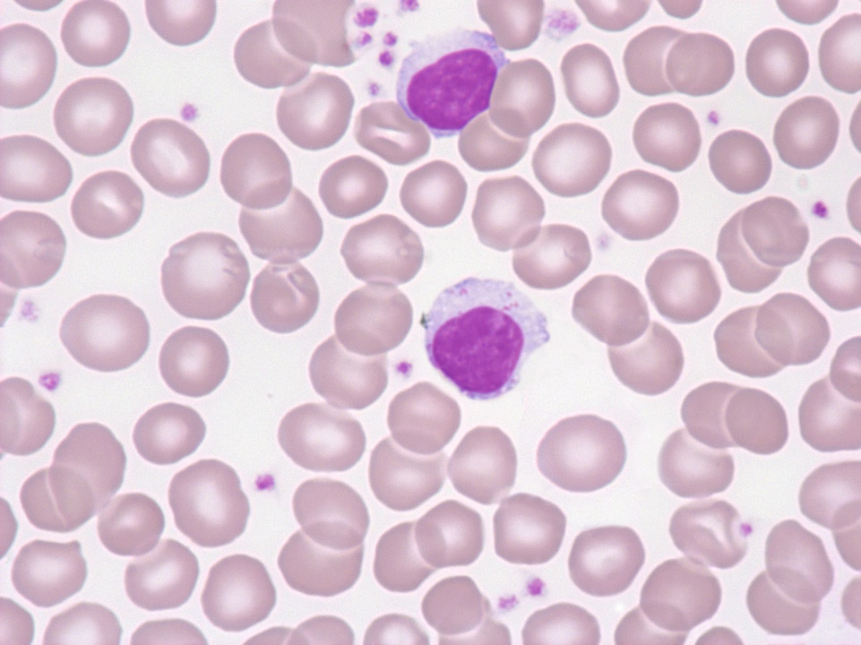
This patient still has a normal white blood cell concentration of 9,500/μL, but a severely reduced granulocyte count (5%, absolute: 425/μL). Red blood cell count and platelets are inconspicuous. Diagnosis: T-cell large granular lymphocytic (LGL) leukaemia.
<p>This patient still has a normal white blood cell concentration of 9,500/μL, but a severely reduced granulocyte count (5%, absolute: 425/μL). Red blood cell count and platelets are inconspicuous. Diagnosis: T-cell large granular lymphocytic (LGL) leukaemia.</p>
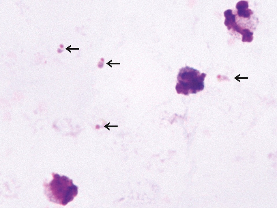
A 'thick blood film' is prepared by spreading a drop of blood on a slide in a way that approximately 20 layers of red blood cells are on top of each other. The slide is then left to dry and subsequently treated with Giemsa solution to lyse the red blood cells. This process leads to a higher density of the malaria pathogens (here: trophozoites of Plasmodium falciparum ->) on the slide so that they can be detected more easily and quickly. The large nuclei are debris from lysed white blood cells.
<p>A 'thick blood film' is prepared by spreading a drop of blood on a slide in a way that approximately 20 layers of red blood cells are on top of each other. The slide is then left to dry and subsequently treated with Giemsa solution to lyse the red blood cells. This process leads to a higher density of the malaria pathogens (here: trophozoites of Plasmodium falciparum ->) on the slide so that they can be detected more easily and quickly. The large nuclei are debris from lysed white blood cells. </p>
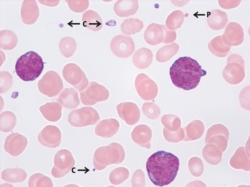
Thrombocytopenia and detection of cytoplasm fragments from monoblasts in AML-M5A. Due to their similar size the cytoplasm fragments can produce a falsely elevated platelet count in impedance measurement on certain haematology analysers. Here the initial count was 32,000/μL. The correct platelet value was obtained from the optical channel; it was 7,000/μL. Cytoplasm fragments (C) and a platelet (P) are marked.
<p>Thrombocytopenia and detection of cytoplasm fragments from monoblasts in AML-M5A. Due to their similar size the cytoplasm fragments can produce a falsely elevated platelet count in impedance measurement on certain haematology analysers. Here the initial count was 32,000/μL. The correct platelet value was obtained from the optical channel; it was 7,000/μL. Cytoplasm fragments (C) and a platelet (P) are marked.</p>
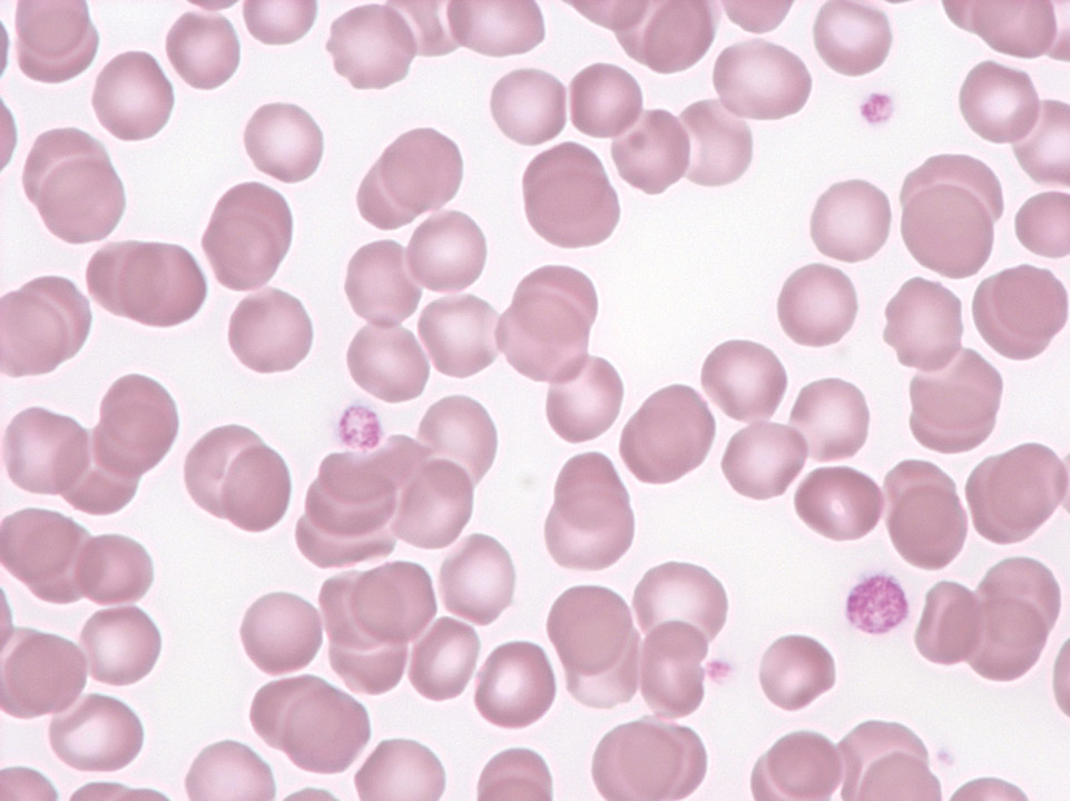
Thrombocytopenia (45,000/μL) and platelet anisocytosis as an indication of idiopathic thrombocytopenic purpura (ITP) in a child after viral infection. IPF (immature platelet fraction) in this case was 18.3%.
<p>Thrombocytopenia (45,000/μL) and platelet anisocytosis as an indication of idiopathic thrombocytopenic purpura (ITP) in a child after viral infection. IPF (immature platelet fraction) in this case was 18.3%.</p>


